| 1) Inspecting the
ECG |
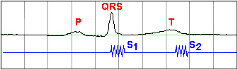
- While displaying Lead II, the
P-wave, the QRS-complex, and the T-wave are
identified. In the program, experiment with different
settings for the time scale and the hardware gain.
|
Time scale: 0.1
secs/division |
Time
scale: 0.5
secs/division | |
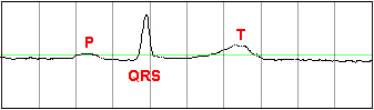
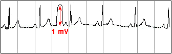
| 2) Calibration |
- While displaying Lead II, the
Cal (calibration) button on the amplifier is momentarily
pressed, giving a pulse that is 1 mV in amplitude.
This pulse can be used to calibrate the amplitude of the ECG
signal. The calibration pulse must not saturate,
therefore one must ensure that the amplifier gain is not set
too high.
|
 | |
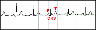
| 3) Identifying Waves and
Intervals |
- The cm/sec switch is set to an
appropriate value for displaying the overall shape of the
P-QRS-T waveform. The various complexes and intervals
are examined. The RR, PR, QRS and QT intervals are
measured, and compared to the normal ranges given in the
table to the right (in seconds).
|
| Interval |
Min |
Max |
| RR |
0.6 |
1.2 |
| PR |
0.12 |
0.20 |
| QRS |
|
0.10 |
| QT |
|
0.42 | |
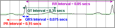
Time scale: 0.1
secs/division | |
| 4) Effect of Lead
Placement |
- The RA electrode is moved from
its position on the wrist to a new position somewhere above
the elbow.
|
 |
 For convenience, the connections of the ECG
electrodes are usually made at the ends of the limbs: at the
wrists and ankles. However, since the limbs act as
conductors, they can be viewed as an extension of the patient
cable lead, and so it makes no difference where the electrodes
are placed along the limb length. For convenience, the connections of the ECG
electrodes are usually made at the ends of the limbs: at the
wrists and ankles. However, since the limbs act as
conductors, they can be viewed as an extension of the patient
cable lead, and so it makes no difference where the electrodes
are placed along the limb length.
|
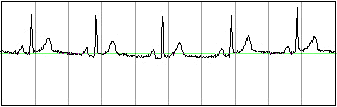
- After returning the RA electrode
to its original position, the subject extends the right arm
outwards and holds it horizontally in mid-air away from the
body.
|
 |
| The above ECG trace appears very noisy, because
the recording is also picking up the EMG activity from the
muscles used in extending the arm
outwards. | |
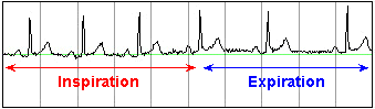
| 5) Effect of
Respiration |
- The subject takes a deep slow
breath, and then exhales slowly (inhaling for 5 seconds, and
exhaling for five seconds).
|
 |
|
In sinus arrhythmia, the
heart rate varies with the phase of respiration. The
heart rate typically increases during inspiration and
decreases during expiration. Therefore, as observed, the
R-R interval is longer during expiration. These changes
are mediated through vagal reflexes. Sinus arrhythmia is
more common in young healthy
athletes. | |
|
|
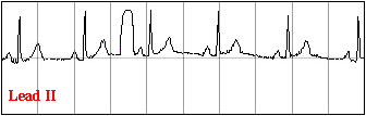
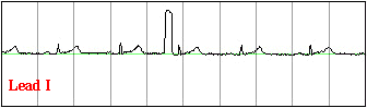
| 7) Changes in Morphology with
Leads |
- The lead selector is switched to
lead I. The group should describe the changes seen
with respect to lead II, and attempt to explain
them.
|
- The lead selector is switched to
lead III. The group should describe the changes seen
with respect to lead II, and attempt to explain
them.
|
 |
 |
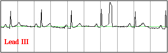 |
| Recall that the R wave is due to the activation
(depolarization) of the major portion of the ventricles.
From the sample data above, it is evident that the lead whose
axis is most parallel to the direction of the
subject's ventricular depolarization is lead II. (The R wave
is largest in lead II.) The R wave is very small in
lead I. We can therefore conclude that for this subject
the direction of ventricular depolarization is more close to
being perpendicular to lead
I. | | |